File:IPLab6RA2.jpg
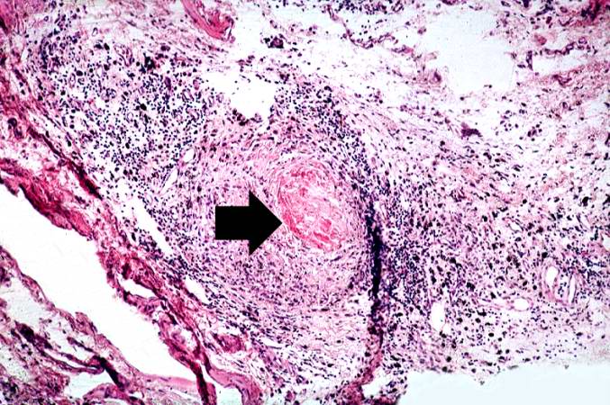
Revision as of 18:06, 19 August 2013 by Seung Park (talk | contribs) (This is a medium-power photomicrograph of the joint capsule surrounding the metacarpal joints. Note the thickening of the capsule and the focal accumulation of inflammatory cells surrounding a central area of fibrinoid necrosis (arrow).)
No higher resolution available.
IPLab6RA2.jpg (677 × 450 pixels, file size: 97 KB, MIME type: image/jpeg)
This is a medium-power photomicrograph of the joint capsule surrounding the metacarpal joints. Note the thickening of the capsule and the focal accumulation of inflammatory cells surrounding a central area of fibrinoid necrosis (arrow).
File history
Click on a date/time to view the file as it appeared at that time.
| Date/Time | Thumbnail | Dimensions | User | Comment | |
|---|---|---|---|---|---|
| current | 18:06, 19 August 2013 | | 677 × 450 (97 KB) | Seung Park (talk | contribs) | This is a medium-power photomicrograph of the joint capsule surrounding the metacarpal joints. Note the thickening of the capsule and the focal accumulation of inflammatory cells surrounding a central area of fibrinoid necrosis (arrow). |
- You cannot overwrite this file.
File usage
There are no pages that link to this file.