File:IPLab4Thrombosis7.jpg
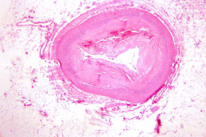
In this low-power photomicrograph of another coronary artery from this patient, a mural thrombus has undergone re-organization. The mural thrombus has been invaded by the in-growth of fibroblasts and small blood vessels from the wall of the artery. The thrombotic material has been phagocytosed and removed by macrophages and is replaced by fibrous connective tissue and blood vessels. This re-organized thrombus still compromises the lumen of this vessel.
Mural thrombosis is the formation of multiple thrombi along an injured endocardial wall.
A thrombus is a solid mass resulting from the aggregation of blood constituents within the vascular system.
File history
Click on a date/time to view the file as it appeared at that time.
| Date/Time | Thumbnail | Dimensions | User | Comment | |
|---|---|---|---|---|---|
| current | 16:40, 19 August 2013 | | 676 × 450 (50 KB) | Seung Park (talk | contribs) | In this low-power photomicrograph of another coronary artery from this patient, a mural thrombus has undergone re-organization. The mural thrombus has been invaded by the in-growth of fibroblasts and small blood vessels from the wall of the artery. The... |
- You cannot overwrite this file.
File usage
The following page links to this file: