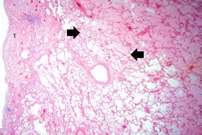
File:IPLab4PulmonaryCongestion5.jpg
Revision as of 16:08, 19 August 2013 by Seung Park (talk | contribs) (This is a higher-power photomicrograph of lung. The edema fluid within the alveoli is visible at this higher magnification (arrows). The thickened pleura (1) is on the left.)
No higher resolution available.
IPLab4PulmonaryCongestion5.jpg (675 × 450 pixels, file size: 63 KB, MIME type: image/jpeg)
This is a higher-power photomicrograph of lung. The edema fluid within the alveoli is visible at this higher magnification (arrows). The thickened pleura (1) is on the left.
File history
Click on a date/time to view the file as it appeared at that time.
| Date/Time | Thumbnail | Dimensions | User | Comment | |
|---|---|---|---|---|---|
| current | 16:08, 19 August 2013 | | 675 × 450 (63 KB) | Seung Park (talk | contribs) | This is a higher-power photomicrograph of lung. The edema fluid within the alveoli is visible at this higher magnification (arrows). The thickened pleura (1) is on the left. |
- You cannot overwrite this file.
File usage
The following page links to this file: