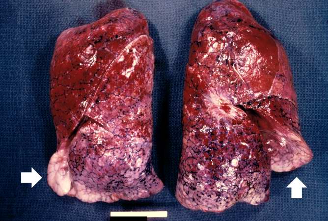
File:IPLab4PulmonaryCongestion1.jpg
Revision as of 16:06, 19 August 2013 by Seung Park (talk | contribs) (This is a gross photograph of lungs that are distended and red. The reddish coloration of the tissue is due to congestion. Some normal pink lung tissue is seen at the edges of the lungs (arrows).)
No higher resolution available.
IPLab4PulmonaryCongestion1.jpg (668 × 450 pixels, file size: 64 KB, MIME type: image/jpeg)
This is a gross photograph of lungs that are distended and red. The reddish coloration of the tissue is due to congestion. Some normal pink lung tissue is seen at the edges of the lungs (arrows).
File history
Click on a date/time to view the file as it appeared at that time.
| Date/Time | Thumbnail | Dimensions | User | Comment | |
|---|---|---|---|---|---|
| current | 16:06, 19 August 2013 | | 668 × 450 (64 KB) | Seung Park (talk | contribs) | This is a gross photograph of lungs that are distended and red. The reddish coloration of the tissue is due to congestion. Some normal pink lung tissue is seen at the edges of the lungs (arrows). |
- You cannot overwrite this file.
File usage
The following page links to this file: