File:IPLab3BrainInfarction7.jpg
Revision as of 04:23, 19 August 2013 by Seung Park (talk | contribs) (This is a photomicrograph of the edge of the infarct. The macrophages that are full of brain tissue (“gitter cells”) are at the top of the image (arrows) and the brain parenchyma containing gemistocytic astrocytes is at the bottom.)
No higher resolution available.
IPLab3BrainInfarction7.jpg (680 × 450 pixels, file size: 50 KB, MIME type: image/jpeg)
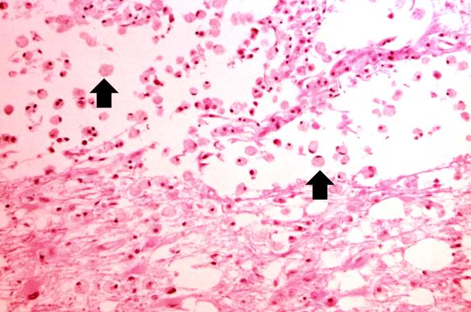
This is a photomicrograph of the edge of the infarct. The macrophages that are full of brain tissue (“gitter cells”) are at the top of the image (arrows) and the brain parenchyma containing gemistocytic astrocytes is at the bottom.
Gemistocytic astrocytes (gemistocytes) comprise a form of activated astrocyte in which the cell body becomes round and swollen, the nucleus assumes an eccentric position, and the cytoplasm changes to an easily visible bright pink color.
File history
Click on a date/time to view the file as it appeared at that time.
| Date/Time | Thumbnail | Dimensions | User | Comment | |
|---|---|---|---|---|---|
| current | 04:23, 19 August 2013 | | 680 × 450 (50 KB) | Seung Park (talk | contribs) | This is a photomicrograph of the edge of the infarct. The macrophages that are full of brain tissue (“gitter cells”) are at the top of the image (arrows) and the brain parenchyma containing gemistocytic astrocytes is at the bottom. |
- You cannot overwrite this file.
File usage
There are no pages that link to this file.