File:IPLab3ChronicPepticUlcer8.jpg
Revision as of 04:16, 19 August 2013 by Seung Park (talk | contribs) (This low-power photomicrograph demonstrates the healing reaction in the base of this ulcer. The base of the ulcer is at the left-hand side of the image and the serosal surface is at the right. Note the fibrous connective tissue within the wall of the s...)
No higher resolution available.
IPLab3ChronicPepticUlcer8.jpg (660 × 450 pixels, file size: 50 KB, MIME type: image/jpeg)
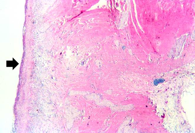
This low-power photomicrograph demonstrates the healing reaction in the base of this ulcer. The base of the ulcer is at the left-hand side of the image and the serosal surface is at the right. Note the fibrous connective tissue within the wall of the stomach and the layer of inflammatory exudate on the surface of the ulcer (arrow).
File history
Click on a date/time to view the file as it appeared at that time.
| Date/Time | Thumbnail | Dimensions | User | Comment | |
|---|---|---|---|---|---|
| current | 04:16, 19 August 2013 | | 660 × 450 (50 KB) | Seung Park (talk | contribs) | This low-power photomicrograph demonstrates the healing reaction in the base of this ulcer. The base of the ulcer is at the left-hand side of the image and the serosal surface is at the right. Note the fibrous connective tissue within the wall of the s... |
- You cannot overwrite this file.
File usage
There are no pages that link to this file.