File:IPLab3Tuberculosis3.jpg
Revision as of 03:39, 19 August 2013 by Seung Park (talk | contribs) (This is a photomicrograph of a tuberculosis granuloma. Note the central core of caseation necrosis (1) encircled by a rim of epithelioid macrophages and lymphocytes (2). Langhans’ type multinucleated giant cells are also present although they are dif...)
No higher resolution available.
IPLab3Tuberculosis3.jpg (683 × 450 pixels, file size: 77 KB, MIME type: image/jpeg)
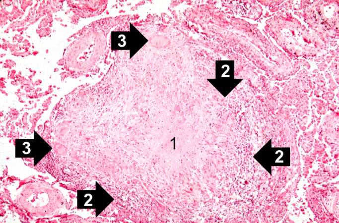
This is a photomicrograph of a tuberculosis granuloma. Note the central core of caseation necrosis (1) encircled by a rim of epithelioid macrophages and lymphocytes (2). Langhans’ type multinucleated giant cells are also present although they are difficult to see at this power (3).
File history
Click on a date/time to view the file as it appeared at that time.
| Date/Time | Thumbnail | Dimensions | User | Comment | |
|---|---|---|---|---|---|
| current | 03:39, 19 August 2013 | | 683 × 450 (77 KB) | Seung Park (talk | contribs) | This is a photomicrograph of a tuberculosis granuloma. Note the central core of caseation necrosis (1) encircled by a rim of epithelioid macrophages and lymphocytes (2). Langhans’ type multinucleated giant cells are also present although they are dif... |
- You cannot overwrite this file.
File usage
The following page links to this file: