No higher resolution available.
IPLab1LungAbscess8.jpg (679 × 450 pixels, file size: 85 KB, MIME type: image/jpeg)
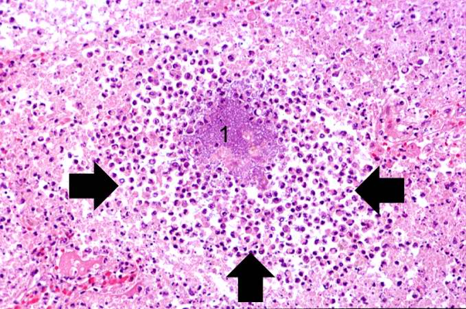
A high-power photomicrograph of lung from this case demonstrates a small abscess full of inflammatory cells (primarily neutrophils) (arrows). There is a bacterial colony in the center of this abscess (1).
An abscess is a collection of pus (white blood cells) within a cavity formed by disintegrated tissue.
File history
Click on a date/time to view the file as it appeared at that time.
| Date/Time | Thumbnail | Dimensions | User | Comment | |
|---|---|---|---|---|---|
| current | 16:15, 15 August 2013 | | 679 × 450 (85 KB) | Seung Park (talk | contribs) |
- You cannot overwrite this file.
File usage
The following page links to this file: