File:IPLab12Mesothelioma7.jpg
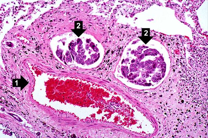
Revision as of 05:33, 21 August 2013 by Seung Park (talk | contribs) (In this higher-power photomicrograph there is a blood vessel (1) and adjacent lymphatics that contain tumor cells (2). There are also accumulations of brown material adjacent to these vessels.)
No higher resolution available.
IPLab12Mesothelioma7.jpg (677 × 450 pixels, file size: 100 KB, MIME type: image/jpeg)
In this higher-power photomicrograph there is a blood vessel (1) and adjacent lymphatics that contain tumor cells (2). There are also accumulations of brown material adjacent to these vessels.
File history
Click on a date/time to view the file as it appeared at that time.
| Date/Time | Thumbnail | Dimensions | User | Comment | |
|---|---|---|---|---|---|
| current | 05:33, 21 August 2013 | | 677 × 450 (100 KB) | Seung Park (talk | contribs) | In this higher-power photomicrograph there is a blood vessel (1) and adjacent lymphatics that contain tumor cells (2). There are also accumulations of brown material adjacent to these vessels. |
- You cannot overwrite this file.
File usage
The following page links to this file: