File:IPLab12Mesothelioma5.jpg
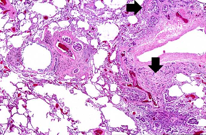
Revision as of 05:33, 21 August 2013 by Seung Park (talk | contribs) (This higher-power photomicrograph of left lung shows areas of consolidation and fibrosis (arrows). Also note that in many of these areas there are clusters of blue cells.)
No higher resolution available.
IPLab12Mesothelioma5.jpg (686 × 450 pixels, file size: 99 KB, MIME type: image/jpeg)
This higher-power photomicrograph of left lung shows areas of consolidation and fibrosis (arrows). Also note that in many of these areas there are clusters of blue cells.
File history
Click on a date/time to view the file as it appeared at that time.
| Date/Time | Thumbnail | Dimensions | User | Comment | |
|---|---|---|---|---|---|
| current | 05:33, 21 August 2013 | | 686 × 450 (99 KB) | Seung Park (talk | contribs) | This higher-power photomicrograph of left lung shows areas of consolidation and fibrosis (arrows). Also note that in many of these areas there are clusters of blue cells. |
- You cannot overwrite this file.
File usage
The following page links to this file: