File:IPLab11Cysticercosis2.jpg
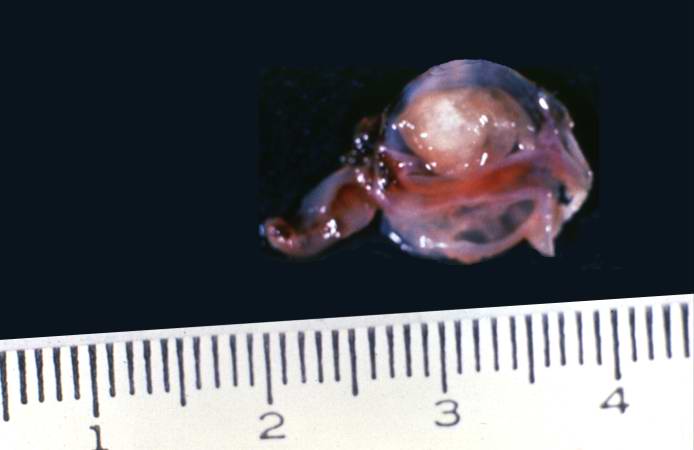
Revision as of 05:03, 21 August 2013 by Seung Park (talk | contribs) (This is a photograph of the cyst that was surgically removed. The cyst is filled with a clear fluid and contains a scolex.)
No higher resolution available.
IPLab11Cysticercosis2.jpg (694 × 450 pixels, file size: 20 KB, MIME type: image/jpeg)
This is a photograph of the cyst that was surgically removed. The cyst is filled with a clear fluid and contains a scolex.
File history
Click on a date/time to view the file as it appeared at that time.
| Date/Time | Thumbnail | Dimensions | User | Comment | |
|---|---|---|---|---|---|
| current | 05:03, 21 August 2013 | | 694 × 450 (20 KB) | Seung Park (talk | contribs) | This is a photograph of the cyst that was surgically removed. The cyst is filled with a clear fluid and contains a scolex. |
- You cannot overwrite this file.
File usage
The following page links to this file: