File:IPLab11Leishmaniasis4.jpg
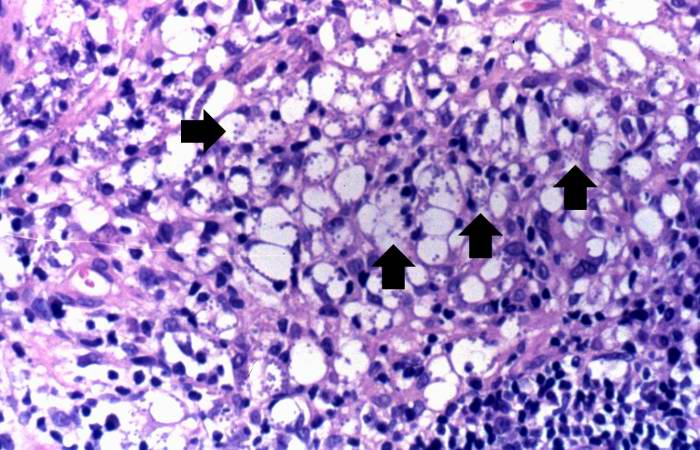
Revision as of 04:58, 21 August 2013 by Seung Park (talk | contribs) (This high-power photomicrograph of the biopsy specimen shows more clearly the heavily infiltrate of inflammatory cells. Note the small blue structures inside the inflammatory cells (arrows).)
No higher resolution available.
IPLab11Leishmaniasis4.jpg (700 × 450 pixels, file size: 70 KB, MIME type: image/jpeg)
This high-power photomicrograph of the biopsy specimen shows more clearly the heavily infiltrate of inflammatory cells. Note the small blue structures inside the inflammatory cells (arrows).
An infiltrate is an accumulation of cells in the lung parenchyma--this is a sign of pneumonia.
File history
Click on a date/time to view the file as it appeared at that time.
| Date/Time | Thumbnail | Dimensions | User | Comment | |
|---|---|---|---|---|---|
| current | 04:58, 21 August 2013 | | 700 × 450 (70 KB) | Seung Park (talk | contribs) | This high-power photomicrograph of the biopsy specimen shows more clearly the heavily infiltrate of inflammatory cells. Note the small blue structures inside the inflammatory cells (arrows). |
- You cannot overwrite this file.
File usage
The following page links to this file: