File:IPLab10Mucor6.jpg
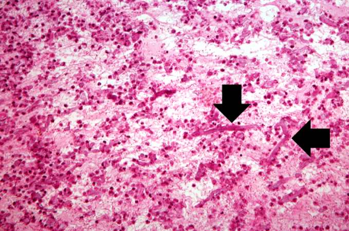
Revision as of 04:20, 21 August 2013 by Seung Park (talk | contribs) (This medium-power photomicrograph shows the thrombus stained to outline the Mucor organisms (arrows). Note again the ribbon-like morphology and the wide-angle branching.)
No higher resolution available.
IPLab10Mucor6.jpg (679 × 450 pixels, file size: 76 KB, MIME type: image/jpeg)
This medium-power photomicrograph shows the thrombus stained to outline the Mucor organisms (arrows). Note again the ribbon-like morphology and the wide-angle branching.
A thrombus is a solid mass resulting from the aggregation of blood constituents within the vascular system.
File history
Click on a date/time to view the file as it appeared at that time.
| Date/Time | Thumbnail | Dimensions | User | Comment | |
|---|---|---|---|---|---|
| current | 04:20, 21 August 2013 | | 679 × 450 (76 KB) | Seung Park (talk | contribs) | This medium-power photomicrograph shows the thrombus stained to outline the Mucor organisms (arrows). Note again the ribbon-like morphology and the wide-angle branching. |
- You cannot overwrite this file.
File usage
The following page links to this file: