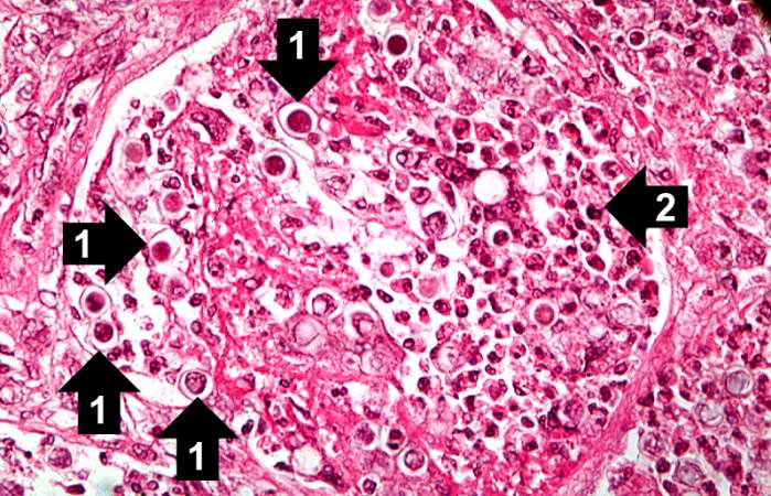
File:IPLab10Blasto7.jpg
Revision as of 04:16, 21 August 2013 by Seung Park (talk | contribs) (This high-power photomicrograph shows an alveolus filled with numerous round bodies up to 25 mm in diameter. Some of these double-contour bodies (1) have a dense center and a clear halo. These are the Blastomyces organisms. The typical B. dermatitides ...)
No higher resolution available.
IPLab10Blasto7.jpg (699 × 450 pixels, file size: 90 KB, MIME type: image/jpeg)
This high-power photomicrograph shows an alveolus filled with numerous round bodies up to 25 mm in diameter. Some of these double-contour bodies (1) have a dense center and a clear halo. These are the Blastomyces organisms. The typical B. dermatitides organism is smoothly-outlined with a central, densely basophilic cytoplasm surrounded by a clear halo. When stained with hematoxylin and eosin, the organism is outlined by a relatively thick cell wall. There are also numerous inflammatory cells (2) in the alveolus--neutrophils, lymphocytes and macrophages--which produce a pyogranulomatous inflammatory reaction.
File history
Click on a date/time to view the file as it appeared at that time.
| Date/Time | Thumbnail | Dimensions | User | Comment | |
|---|---|---|---|---|---|
| current | 04:16, 21 August 2013 | | 699 × 450 (90 KB) | Seung Park (talk | contribs) | This high-power photomicrograph shows an alveolus filled with numerous round bodies up to 25 mm in diameter. Some of these double-contour bodies (1) have a dense center and a clear halo. These are the Blastomyces organisms. The typical B. dermatitides ... |
- You cannot overwrite this file.
File usage
The following page links to this file: