File:IPLab10Histo6.jpg
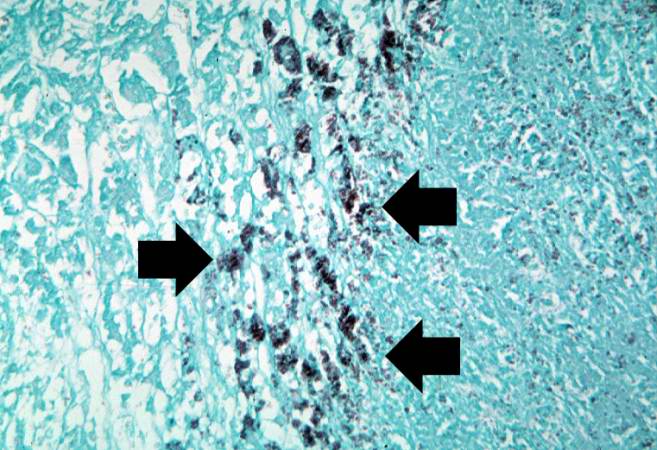
Revision as of 04:07, 21 August 2013 by Seung Park (talk | contribs) (This is a high-power photomicrograph of the same section of tissue as the previous slide. This section, however, has been stained with methenamine silver which causes the Histoplasma organisms to stain black (arrows).)
No higher resolution available.
IPLab10Histo6.jpg (657 × 450 pixels, file size: 67 KB, MIME type: image/jpeg)
This is a high-power photomicrograph of the same section of tissue as the previous slide. This section, however, has been stained with methenamine silver which causes the Histoplasma organisms to stain black (arrows).
File history
Click on a date/time to view the file as it appeared at that time.
| Date/Time | Thumbnail | Dimensions | User | Comment | |
|---|---|---|---|---|---|
| current | 04:07, 21 August 2013 | | 657 × 450 (67 KB) | Seung Park (talk | contribs) | This is a high-power photomicrograph of the same section of tissue as the previous slide. This section, however, has been stained with methenamine silver which causes the Histoplasma organisms to stain black (arrows). |
- You cannot overwrite this file.
File usage
The following page links to this file: