File:IPLab10Candidiasis1.jpg
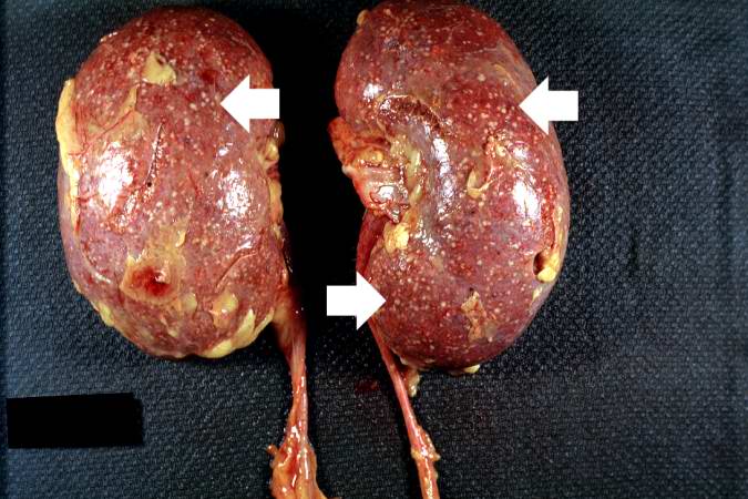
Revision as of 04:02, 21 August 2013 by Seung Park (talk | contribs) (This autopsy photograph of the kidneys demonstrates the multifocal punctate lesions visible on the serosal surface (arrows). Don't confuse these small yellow punctate lesions with the fat that is adherent to the renal capsule.)
No higher resolution available.
IPLab10Candidiasis1.jpg (675 × 450 pixels, file size: 64 KB, MIME type: image/jpeg)
This autopsy photograph of the kidneys demonstrates the multifocal punctate lesions visible on the serosal surface (arrows). Don't confuse these small yellow punctate lesions with the fat that is adherent to the renal capsule.
File history
Click on a date/time to view the file as it appeared at that time.
| Date/Time | Thumbnail | Dimensions | User | Comment | |
|---|---|---|---|---|---|
| current | 04:02, 21 August 2013 | | 675 × 450 (64 KB) | Seung Park (talk | contribs) | This autopsy photograph of the kidneys demonstrates the multifocal punctate lesions visible on the serosal surface (arrows). Don't confuse these small yellow punctate lesions with the fat that is adherent to the renal capsule. |
- You cannot overwrite this file.
File usage
The following page links to this file: