Difference between revisions of "File:IPLab2Metaplasia2.jpg"
(his high-power photomicrograph demonstrates the transitional epithelium lining the renal calyx (1) and the junction (transition zone) to a thicker hyperplastic epithelium (2). Note the inflammatory cells and increased vascular response in the stromal t...) |
|||
| Line 1: | Line 1: | ||
| − | + | This high-power photomicrograph demonstrates the transitional epithelium lining the renal calyx (1) and the junction (transition zone) to a thicker hyperplastic epithelium (2). Note the inflammatory cells and increased vascular response in the stromal tissue (3) lying beneath the normal transitional epithelium. | |
Latest revision as of 15:48, 19 August 2013
This high-power photomicrograph demonstrates the transitional epithelium lining the renal calyx (1) and the junction (transition zone) to a thicker hyperplastic epithelium (2). Note the inflammatory cells and increased vascular response in the stromal tissue (3) lying beneath the normal transitional epithelium.
File history
Click on a date/time to view the file as it appeared at that time.
| Date/Time | Thumbnail | Dimensions | User | Comment | |
|---|---|---|---|---|---|
| current | 15:40, 19 August 2013 | 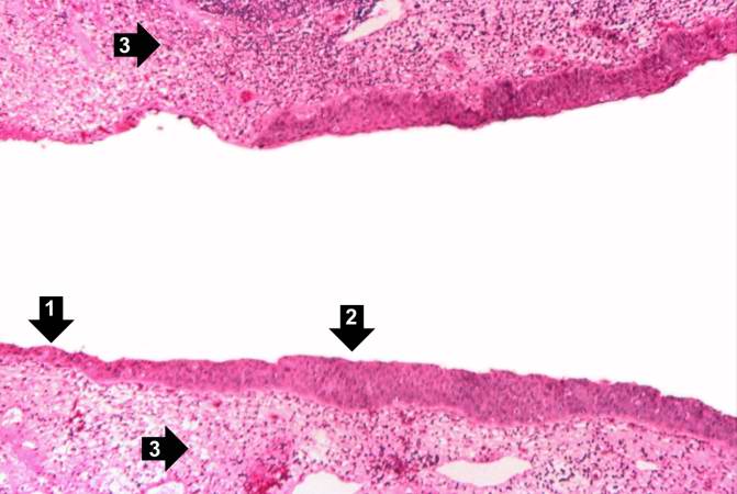 | 671 × 450 (41 KB) | Peter Anderson (talk | contribs) | his high-power photomicrograph demonstrates the transitional epithelium lining the renal calyx (1) and the junction (transition zone) to a thicker hyperplastic epithelium (2). Note the inflammatory cells and increased vascular response in the stromal t... |
- You cannot overwrite this file.
File usage
There are no pages that link to this file.