IPLab:Lab 13:Meningococcemia: Difference between revisions
Seung Park (talk | contribs) No edit summary |
Seung Park (talk | contribs) No edit summary |
||
| Line 21: | Line 21: | ||
* <spoiler text="What is the source of Neisseria meningitides?">Man is the only host. The organism lives in the nasopharynges of apparently unaffected individuals.</spoiler> | * <spoiler text="What is the source of Neisseria meningitides?">Man is the only host. The organism lives in the nasopharynges of apparently unaffected individuals.</spoiler> | ||
* <spoiler text="What is the favorite site of growth of Neisseria meningitides during clinical illness?">Cerebrospinal fluid is the favorite site of growth for the bacteria and meningitis is the most common illness. Other sites include heart valves, pericardium, joints, and the lungs.</spoiler> | * <spoiler text="What is the favorite site of growth of Neisseria meningitides during clinical illness?">Cerebrospinal fluid is the favorite site of growth for the bacteria and meningitis is the most common illness. Other sites include heart valves, pericardium, joints, and the lungs.</spoiler> | ||
== Additional Resources == | |||
=== Reference === | |||
=== Journal Articles === | |||
=== Images === | |||
== Related IPLab Cases == | |||
{{IPLab 13}} | {{IPLab 13}} | ||
[[Category: IPLab:Lab 13]] | [[Category: IPLab:Lab 13]] | ||
Revision as of 23:28, 22 August 2013
Clinical Summary
This 20-month-old black female was active and without complaint until 4 p.m. on the evening prior to her death, when according to her mother she acted as if she did not feel well. The mother reported that the child had felt warm at bedtime (8 p.m.) so she had given her some acetaminophen. At midnight, the child was given another dose of acetaminophen. The child slept with her mother that night who reported last hearing her daughter make a sound at approximately 7:30 a.m. The mother checked in on her daughter at 8:45 a.m. and found her to be unresponsive. The girl was dead when paramedics arrived. Her past medical history was unremarkable and there had been no recent illness among other family members.
Autopsy Findings
External examination revealed multiple purpuric and ecchymotic cutaneous lesions, most notable in the inguinal areas. Microscopic examination of these skin lesions revealed thrombosis and rupture of small dermal vessels. There were scattered visceral petechial hemorrhages and both adrenal glands were grossly hemorrhagic. There was no gross or microscopic evidence of meningitis. However, a Gram stain of cerebrospinal fluid showed Gram-negative diplococci. Cultures were positive for Neisseria meningitides.
Images
-
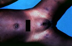 In this gross photograph from the autopsy, note the areas of hemorrhage in the inguinal region.
 -
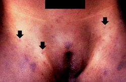 This is a closer view of the inguinal region taken at autopsy. The areas of hemorrhage include purpura and petechiae (arrows).
 -
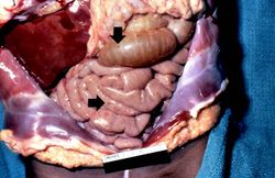 In this gross photograph of the abdomen taken at the autopsy, there are petechial hemorrhages on the viscera (arrows).
 -
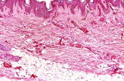 This photomicrograph of the skin shows thrombi and fibrin clots in small vessels in the dermis. This is indicative of the endothelial damage caused by the Neisseria meningitidis endotoxin. This endotoxin-induced damage to the endothelium of small blood vessels throughout the body results in widespread petechiae and purpura.
 -
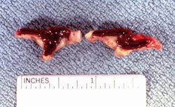 This is a gross photograph of cross sections through the adrenal glands from this case. Both adrenal glands are markedly hemorrhagic.
 -
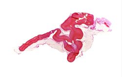 This is a low-power photomicrograph of the adrenal gland from this case. Note that the entire gland is hemorrhagic.
 -
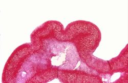 This higher-power photomicrograph of the adrenal gland from this case provides an example of hemorrhagic necrosis.
 -
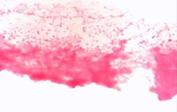 This is a higher-power photomicrograph of a smear of cerebrospinal fluid taken at autopsy. Note the Gram-negative cocci in this smear, indicative of N. meningitidis.


Study Questions
Additional Resources
Reference
Journal Articles
Images
Related IPLab Cases
| |||||