IPLab:Lab 9:Diphtheria: Difference between revisions
Jump to navigation
Jump to search
Seung Park (talk | contribs) No edit summary |
|||
| (One intermediate revision by the same user not shown) | |||
| Line 2: | Line 2: | ||
This 4-year-old black female had an upper respiratory infection and a sore throat with increasing difficulty in breathing. Membranous exudate over one tonsil led to a working diagnosis of diphtheria, and the child was admitted. On the day of her admission, the child developed signs of respiratory tract obstruction and a tracheotomy was performed. However, the procedure was unable to establish a patent airway and the child died. | This 4-year-old black female had an upper respiratory infection and a sore throat with increasing difficulty in breathing. Membranous exudate over one tonsil led to a working diagnosis of diphtheria, and the child was admitted. On the day of her admission, the child developed signs of respiratory tract obstruction and a tracheotomy was performed. However, the procedure was unable to establish a patent airway and the child died. | ||
At autopsy, a dense grayish pink membrane extended from both tonsils to the mid-trachea. The lungs were edematous and showed signs of pneumonia. | |||
At autopsy, a dense grayish pink membrane extended from both tonsils to the mid-trachea. The lungs were edematous and showed signs of pneumonia. | |||
== Images == | == Images == | ||
Latest revision as of 16:43, 9 July 2020
Clinical Summary
This 4-year-old black female had an upper respiratory infection and a sore throat with increasing difficulty in breathing. Membranous exudate over one tonsil led to a working diagnosis of diphtheria, and the child was admitted. On the day of her admission, the child developed signs of respiratory tract obstruction and a tracheotomy was performed. However, the procedure was unable to establish a patent airway and the child died.
At autopsy, a dense grayish pink membrane extended from both tonsils to the mid-trachea. The lungs were edematous and showed signs of pneumonia.
Images
-
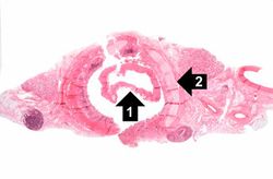 This is a low-power photomicrograph of the trachea with the diphtheritic membrane (1), which has pulled away from the tracheal lining during histological processing. Note the tracheal cartilage (2) present in this section.
 -
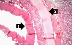 This is a higher-power photomicrograph of trachea with the diphtheritic membrane (1). Even though, the main part of the membrane has pulled away from the tracheal lining during histological processing, in this section part of the membrane is still loosely attached. Once again, note the tracheal cartilage (2).
 -
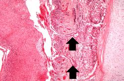 This is an even higher-power photomicrograph of the tracheal mucosa and the diphtheritic membrane. The mucosal surface of the trachea is ulcerated (total loss of epithelial cells) and the only remaining epithelial cells are found in the glands (arrows). The diphtheritic membrane consists of fibrin and inflammatory cells, most of which are dead.
 -
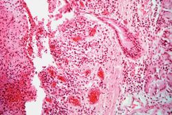 In this higher-power photomicrograph of the tissue from the previous image, the ulcerated tracheal mucosa and the diphtheritic membrane are more clearly seen. Although difficult to make out at this magnification, most of the cells in this inflammatory exudate are neutrophils.


Virtual Microscopy
Study Questions
Additional Resources
Reference
- eMedicine Medical Library: Diphtheria
- eMedicine Medical Library: Pediatric Pneumonia
- eMedicine Medical Library: Corynebacterium Infections
- Merck Manual: Diphtheria
Journal Articles
- Zakikhany K, Efstratiou A. Diphtheria in Europe: current problems and new challenges. Future Microbiol 2012 May;7(5):595-607.
Images
| |||||