IPLab:Lab 5:Neurofibromatosis: Difference between revisions
Jump to navigation
Jump to search
Seung Park (talk | contribs) No edit summary |
|||
| (6 intermediate revisions by 2 users not shown) | |||
| Line 1: | Line 1: | ||
== Clinical Summary == | == Clinical Summary == | ||
A 45-year-old divorced white male came to the emergency room with severe hepatic cirrhosis and aspiration pneumonia. Shortly after admission he developed cardiac arrhythmias and died. Significant past history included alcohol abuse, cirrhosis, and neurofibromatosis. He had no family history of neurofibromatosis, but his condition was diagnosed at age 17 when he developed neurofibromas along the lateral chest wall. There was no history of continued follow-up after this initial diagnosis. | |||
The patient was covered with variably sized subcutaneous nodules ranging from 0.5 to 2.5 cm in diameter. Other significant findings included micronodular hepatic cirrhosis, ascites (500 ml), and splenomegaly. | |||
The patient was covered with variably sized subcutaneous nodules ranging from 0.5 to 2.5 cm in diameter. Other significant findings included micronodular hepatic cirrhosis | |||
== Images == | == Images == | ||
<gallery heights="250px" widths="250px"> | <gallery heights="250px" widths="250px"> | ||
File: | File:IPLab5Neurofibromatosis1b.JPG|This photograph, taken at autopsy, demonstrates the distribution of neurofibromas on the skin of this patient. | ||
File: | File:IPLab5Neurofibromatosis2b.JPG|This is another view taken at autopsy demonstrating the neurofibromas. Some lesions can be seen as subcutaneous swellings (arrow) and others form pedunculated masses. Most are hyperpigmented. | ||
File: | File:IPLab5Neurofibromatosis3b.JPG|This is a closer view of neurofibromas on the skin. | ||
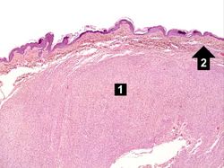
File:IPLab5Neurofibromatosis4.jpg|This is a low-power photomicrograph of a subcutaneous neurofibroma (1). Note the increased pigmentation in the skin (2). | File:IPLab5Neurofibromatosis4.jpg|This is a low-power photomicrograph of a subcutaneous neurofibroma (1). Note the increased pigmentation in the skin (2). | ||
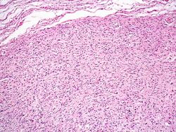
File:IPLab5Neurofibromatosis5.jpg|This is a higher-power photomicrograph of the neurofibroma (1) with the overlying skin (2). | File:IPLab5Neurofibromatosis5.jpg|This is a higher-power photomicrograph of the neurofibroma (1) with the overlying skin (2). | ||
| Line 16: | Line 15: | ||
File:IPLab5Neurofibromatosis8.jpg|This is a high-power photomicrograph of the cells in the neurofibroma. | File:IPLab5Neurofibromatosis8.jpg|This is a high-power photomicrograph of the cells in the neurofibroma. | ||
</gallery> | </gallery> | ||
== Virtual Microscopy == | |||
<peir-vm>IPLab5Neurofibromatosis</peir-vm> | |||
== Study Questions == | == Study Questions == | ||
| Line 22: | Line 24: | ||
* <spoiler text="What are the classic clinical features of neurofibromatosis type 1?">Although protean in its clinical expression, patients with neurofibromatosis usually have multiple neural tumors all over the body, numerous pigmented skin lesions (”café au lait“ spots), and pigmented iris hamartomas (also called Lisch nodules). Other common clinical manifestations include skeletal lesions (scoliosis, bone cysts, thinning of the cortex of long bones) and mild intellectual impairment.</spoiler> | * <spoiler text="What are the classic clinical features of neurofibromatosis type 1?">Although protean in its clinical expression, patients with neurofibromatosis usually have multiple neural tumors all over the body, numerous pigmented skin lesions (”café au lait“ spots), and pigmented iris hamartomas (also called Lisch nodules). Other common clinical manifestations include skeletal lesions (scoliosis, bone cysts, thinning of the cortex of long bones) and mild intellectual impairment.</spoiler> | ||
* <spoiler text="What is the etiology of the coagulopathy seen in this patient?">Liver failure.</spoiler> | * <spoiler text="What is the etiology of the coagulopathy seen in this patient?">Liver failure.</spoiler> | ||
== Additional Resources == | |||
=== Reference === | |||
* [http://emedicine.medscape.com/article/1177266-overview eMedicine Medical Library: Type 1 Neurofibromatosis] | |||
* [http://emedicine.medscape.com/article/1112001-overview eMedicine Medical Library: Dermatologic Manifestations of Neurofibromatosis Type 1] | |||
* [http://www.merckmanuals.com/professional/pediatrics/neurocutaneous_syndromes/neurofibromatosis.html Merck Manual: Neurofibromatosis] | |||
* [http://www.nlm.nih.gov/medlineplus/neurofibromatosis.html Neurofibromatosis: MedlinePlus] | |||
=== Journal Articles === | |||
* Kiliç S, Tezcan I, Sanal O, Ersoy F. [http://www.ncbi.nlm.nih.gov/pubmed/11737751 Common variable immunodeficiency in a patient with neurofibromatosis]. ''Pediatr Int'' 2001 Dec;43(6):691-3. | |||
=== Images === | |||
* [{{SERVER}}/library/index.php?/tags/2145-neurofibromatosis PEIR Digital Library: Neurofibromatosis Images] | |||
* [http://library.med.utah.edu/WebPath/CNSHTML/CNSIDX.html#11 WebPath: CNS Neoplasms] | |||
{{IPLab 5}} | {{IPLab 5}} | ||
[[Category: IPLab:Lab 5]] | [[Category: IPLab:Lab 5]] | ||
Latest revision as of 14:29, 8 July 2020
Clinical Summary
A 45-year-old divorced white male came to the emergency room with severe hepatic cirrhosis and aspiration pneumonia. Shortly after admission he developed cardiac arrhythmias and died. Significant past history included alcohol abuse, cirrhosis, and neurofibromatosis. He had no family history of neurofibromatosis, but his condition was diagnosed at age 17 when he developed neurofibromas along the lateral chest wall. There was no history of continued follow-up after this initial diagnosis.
The patient was covered with variably sized subcutaneous nodules ranging from 0.5 to 2.5 cm in diameter. Other significant findings included micronodular hepatic cirrhosis, ascites (500 ml), and splenomegaly.
Images
-
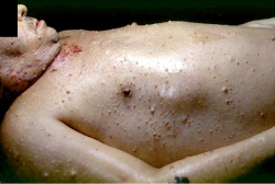 This photograph, taken at autopsy, demonstrates the distribution of neurofibromas on the skin of this patient.
 -
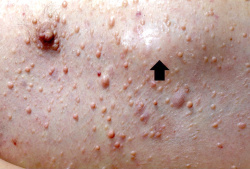 This is another view taken at autopsy demonstrating the neurofibromas. Some lesions can be seen as subcutaneous swellings (arrow) and others form pedunculated masses. Most are hyperpigmented.
 -
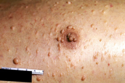 This is a closer view of neurofibromas on the skin.
 -
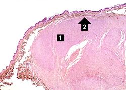 This is a low-power photomicrograph of a subcutaneous neurofibroma (1). Note the increased pigmentation in the skin (2).
 -
 This is a higher-power photomicrograph of the neurofibroma (1) with the overlying skin (2).
 -
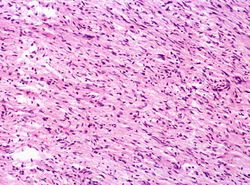
 This is a higher-power photomicrograph of the neurofibroma demonstrating the loose pattern of elongated cells making up the tumor mass.
 -
 This higher-power photomicrograph of the neurofibroma shows more clearly the elongated cells (primarily Schwann cells) that make up this tumor.
 -
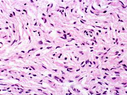 This is a high-power photomicrograph of the cells in the neurofibroma.


Virtual Microscopy
Study Questions
Additional Resources
Reference
- eMedicine Medical Library: Type 1 Neurofibromatosis
- eMedicine Medical Library: Dermatologic Manifestations of Neurofibromatosis Type 1
- Merck Manual: Neurofibromatosis
- Neurofibromatosis: MedlinePlus
Journal Articles
- Kiliç S, Tezcan I, Sanal O, Ersoy F. Common variable immunodeficiency in a patient with neurofibromatosis. Pediatr Int 2001 Dec;43(6):691-3.
Images
| |||||