)
)

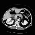
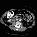
RADIOLOGY: KIDNEY: Case# 33939: Patient is 77 year old female with gross hematuria localized to the right kidney by recent cystoscopy and retrograde pyelogram. 1. Mild right hydronephrosis with an enhancing region seen in the inferior renal pelvis/proximal most ureter which is unusual in appearance. Streaky inflammatory changes are seen surrounding the inferior aspect of the right kidney and proximal ureter. A right ureteral stent is present. These images were compared to a retrograde pyelogram performed which demonstrated marked irregularity of the calyces and pelvis. Tuberculosis should be considered. An unusual presentation of transitional cell carcinoma is also possible. 2. A region of high attenuation is seen within the cortex of the mid portion of the right kidney possibly secondary to retained contrast within a diverticulum. 3. Multiple bilateral simple appearing cysts are seen within the kidneys. 4. A small amount of atelectasis vs. consolidation is seen in the right lower lobe.
- Author
- Peter Anderson
- Posted on
- Thursday 1 August 2013
- Albums
- Visits
- 843
0 comments