)
)
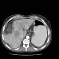
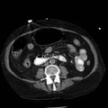
. 2. Collateral vascular pathways include the recanalized umbilical vein along with soft tissue and mesenteric collaterals. 3. Small amount of ascites.")
RADIOLOGY: HEPATOBILIARY: Case# 33795: HEPATOCELLULAR CARCINOMA. 39-year-old female with cirrhosis. Ultrasound examination on the 17th of this month showed two ill-defined lesions in the right lobe of the liver. 1. Small cirrhotic liver with very abnormal perfusion behavior. This may simply be a consequence of the highly abnormal architecture of the liver. Additionally, there is at least one lesion in the right lobe which suggests hepatocellular carcinoma. This lesion involves the surface of the liver and is not an optimum lesion for percutanous biopsy (consensus decision from review of images in CT conference). 2. Collateral vascular pathways include the recanalized umbilical vein along with soft tissue and mesenteric collaterals. 3. Small amount of ascites.
- Author
- Peter Anderson
- Posted on
- Thursday 1 August 2013
- Tags
- hepatobiliary, radiology
- Albums
- Visits
- 684
0 comments