270/452
)
)

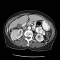
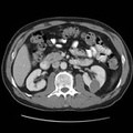
RADIOLOGY: KIDNEY: Case# 33509: RENAL CELL CARCINOMA. The patient is a 62 year old male who has a failing heart and a kidney mass in the posterior aspect of the mid left kidney. 1. A low attenuation mass is seen in the posterior aspect, left mid kidney which enhances following contrast administration and is worrisome for neoplasm. In addition, cysts are seen in the kidneys bilaterally. The left renal vein, inferior vena cava are normal. No significant adenopathy seen. 2. Poorly defined low attenuation area measuring less than 1 cm in the medial segment near the dome of the liver. This may represent volume averaging of adjacent subphrenic fat. Small cysts, metastasis cannot be completely ruled out.
- Author
- Peter Anderson
- Posted on
- Thursday 1 August 2013
- Albums
- Visits
- 771
0 comments