)
)
. 3. Moderate bilateral pleural effusions with small amount of adjacent compressive atelectasis. 4. Small kidneys bilaterally with a 1.5 cm left upper pole cystic lesion which either represents high density cyst or homogeneous mass. When patient has stabilized, pre and suggested to excluded malignancy but even if this is a malignancy it is unlikely to have metastasized at this size. 5. Left medial segment liver lesion has appearance characteristic for hemangioma.")
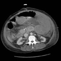
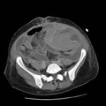
RADIOLOGY: ABDOMEN: Case# 33679: Patient is a 47 year old female on peritoneal dialysis who developed peritonitis and GI bleed with profound hypotension. Patient was too unstable to scan yesterday. 1. Large proximal small bowel intramural and mesenteric hematoma with active contrast extravasation and small bowel obstruction. 2. Multiple extraluminal fluid collections in the abdomen and pelvis consistent with abscesses (greater than 10). 3. Moderate bilateral pleural effusions with small amount of adjacent compressive atelectasis. 4. Small kidneys bilaterally with a 1.5 cm left upper pole cystic lesion which either represents high density cyst or homogeneous mass. When patient has stabilized, pre and suggested to excluded malignancy but even if this is a malignancy it is unlikely to have metastasized at this size. 5. Left medial segment liver lesion has appearance characteristic for hemangioma.
- Author
- Peter Anderson
- Posted on
- Thursday 1 August 2013
- Albums
- Visits
- 979
0 comments