)
)

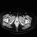
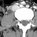
RADIOLOGY: GENITOURINARY: GU: Case# 33032: UVJ STONE W/HYDRONEPHROSIS. The patient is a 21-year-old female with right flank pain. She has a history of leukemia and sarcoma of the breast and has had breast reconstructive surgery on the left. There is moderate hydro-ureteronephrosis on the right down to the level of the ureterovesicle junction. At the ureterovesicle junction, there is a 4 mm high attenuation object within the distal ureter and it is consistent with a ureteral calculus. There is no evidence of intra-abdominal mass or adenopathy. Stone disease is usually idiopathic but may be associated with chronic UTI due to Proteus, hyperparathyroidism, gout, and homocystinuria. Radio-opaque stones such as calcium and some cystine stones may be visualized on plain abdominal films;however, visualizing radiolucent stones requires CT imaging. Radiolucent stones, such as uric acid, xanthine and cystine stones, have a higher attenuation than urine, renal parenchyma and blood clots on CT and thus can be differentiated from other causes of filling defects such a tumor or hematoma. The diagnosis of obstruction and hydronephrosis may be made with or without the use of contrast. It may be easier, however, to distinguish parenchyma from dilated calices with contrast. Extra-renal pelves, dilated calices secondary to post-obtructive atrophy, and parapelvic cysts may mimic changes due to hydronephrosis. On non-contrast-enhanced images, the dilated urine filled calices appear as low attenuation fluid filled structures within the normal or enlarged renal silhouette. On contrast-enhanced supine images, urine typically layers posteriorly. In the case of infection, however, there is "reverse layering" with the high density urine laden with infection rising to the top and the regular urine remaining at the bottom creating the apperance of a fluid level. The affected kidney may show delayed excretion of contrast resulting in a persistent nephrogram. Parenchymal thinning from chronic obstruction may also be evident.
- Author
- Peter Anderson
- Posted on
- Thursday 1 August 2013
- Tags
- Genitourinary, radiology
- Albums
- Visits
- 1170
0 comments