)
)

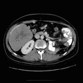
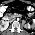
RADIOLOGY: KIDNEY: Case# 33006: RENAL CELL METS. 41 year old black male with history of renal cell carcinoma treated with left radical nephrectomy and x-ray therapy. Evaluate metastatic tumors. Within the liver there is a low attenuation mass measuring 11 x 5 cm adjacent to the posterior aspect of the right hepatic lobe which is probably intrahepatic. Another intrahepatic mass is present within the posterior right lobe measuring 9 x 6 cm. In the left upper quadrant there is a low attenuation mass measuring 10.5 x 9 cm within the peritoneum. A retrosplenic mass is present measuring 5 x 2.5 cm. No definite intraparenchymal lesions are seen within the spleen. Milk of calcium is seen within a normal appearing gallbladder. The pancreas, adrenals, right kidney, and bladder appear within normal limits. The left kidney is surgically absent.
- Author
- Peter Anderson
- Posted on
- Thursday 1 August 2013
- Albums
- Visits
- 1078
0 comments