)
)
. Predisposing factors for colorectal cancer include adenomatous polyps, inherited multiple polyposis syndromes, long-standing ulcerative colitis, close relatives with colon cancer, and a low fiber, high animal fat diet. Colorectal cancer varies in gross presentation according to the region of the colon involved. Carcinoma of the rectosigmoid colon usually presents in an annular manner, often producing early bowel obstruction. Carcinoma of the right colon, however, usually does not obstruct early and frequently presents with iron deficiency anemia secondary to chronic blood loss. Colon cancer spreads by direct extension due to penetration of the colon wall, lymphatic drainage to regional nodes, through the portal system to the liver, and intraperitoneal seeding. Although barium enemas and colonoscopy are the primary methods used to initially diagnose colorectal cancer, CT is an excellent procedure for the detection of recurrence. CT findings of colon cancer include focal lobulated soft tissue mass in the colon, localized thickening of greater than 5 mm of the bowel wall, irregular lumen surface, extension of linear soft tissue densities or discrete mass into pericolic fat or adjacent organs, regional adenopathy, and liver metastases. Soft tissue masses visualized within the peritoneal cavity may signify metastases or recurrence of the cancer. Calcification in the primary tumor can be seen with mucinous adenocarcinoma.")
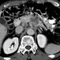
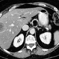
RADIOLOGY: GASTROINTESTINAL: GI: Case# 32888: COLON CANCER METASTASES. The patient is a 62-year-old female metastatic colon carcinoma diagnosed September of 1992 for pre protocol re- evaluation. Comparison is made with a prior CT scan. There are again noted multiple non-calcified pulmonary nodules above lung bases which are increased in size and number. There is again identified low density round lesion in the anterior segment of the right hepatic lobe which previously measured 3.4 x 2.9 cm and now measures 4.0 x 3.0 cm. A second lesion in the lateral left lobe measured 2.8 x 2.5 cm on the previous scan and now measures 2.9 x 2.8 cm. The mesenteric nodal mass measured 1.8 cm and is unchanged. There is evidence of previous right colectomy with transverse ileocolic anastomosis. There is no evidence of local recurrence, retroperitoneal lymph node enlargement, or new hepatic lesions. The spleen, kidneys, adrenals, pancreas, and small and large bowel are otherwise radiographically unremarkable. Colorectal adenocarcinoma is one of the most common neoplasms encountered in the affluent countries of the Western world. The disease exhibits a peak age incidence in the sixth and seventh decades. It is associated with increased serum concentration of carcinoembryonic antigen (CEA). Predisposing factors for colorectal cancer include adenomatous polyps, inherited multiple polyposis syndromes, long-standing ulcerative colitis, close relatives with colon cancer, and a low fiber, high animal fat diet. Colorectal cancer varies in gross presentation according to the region of the colon involved. Carcinoma of the rectosigmoid colon usually presents in an annular manner, often producing early bowel obstruction. Carcinoma of the right colon, however, usually does not obstruct early and frequently presents with iron deficiency anemia secondary to chronic blood loss. Colon cancer spreads by direct extension due to penetration of the colon wall, lymphatic drainage to regional nodes, through the portal system to the liver, and intraperitoneal seeding. Although barium enemas and colonoscopy are the primary methods used to initially diagnose colorectal cancer, CT is an excellent procedure for the detection of recurrence. CT findings of colon cancer include focal lobulated soft tissue mass in the colon, localized thickening of greater than 5 mm of the bowel wall, irregular lumen surface, extension of linear soft tissue densities or discrete mass into pericolic fat or adjacent organs, regional adenopathy, and liver metastases. Soft tissue masses visualized within the peritoneal cavity may signify metastases or recurrence of the cancer. Calcification in the primary tumor can be seen with mucinous adenocarcinoma.
- Author
- Peter Anderson
- Posted on
- Thursday 1 August 2013
- Tags
- gastrointestinal, radiology
- Albums
- Visits
- 795
0 comments