)
)

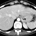
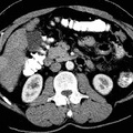
RADIOLOGY: HEPATOBILIARY: Case# 32865: FOCAL NODULAR HYPERPLASIA. 45 year old white female with history of newly identified liver masses at an outside hospital. Liver biopsy demonstrated benign nodular hyperplasia. Four liver lesions are identified. The lesion within the medial segment of the left lobe of the liver measures 3.5x5cm. Inferiorly a separate lesion within the medial segment of the left lobe of the liver measures 8x7cm. A lesion within the lateral segment of the left lobe of the liver measures 2x1.5cm and a separate anterior segment right lobe of the liver lesion measures 1.5x1.5cm. All of these lesions demonstrate low attenuation on non-contrast images. The lesions demonstrate contrast enhancement on the arterial phase and appear isointense to the liver parenchyma on the delayed and portal venous phase images. No other focal liver lesions are identified. No intrahepatic or extrahepatic biliary ductal dilatation is seen.
- Author
- Peter Anderson
- Posted on
- Thursday 1 August 2013
- Tags
- hepatobiliary, radiology
- Albums
- Visits
- 820
0 comments