)
)
 without fever. There is a 14.0 x 9.0 x 10.0 cm low attenuation mass with minimal peripheral enhancement and thick septations associated with its superior aspect. There is minimal associated biliary dilatation. Pyogenic liver abscesses occur predominantly in individuals with other underlying disorders, most commonly biliary tract disease. Abscesses may also occur following hepatic transplantation. Clinical findings are nonspecific with fever and jaundice being the most common. Diagnosis is best made by contrast-enhanced CT or ultrasound. Pyogenic abscesses are usually solitary and often multiloculated with thickened enhancing walls. Gas is present in about 20% of cases. Amoebic abscesses are also solitary lesions. Pyogenic abscesses may be distinguished from amoebic abscesses on the basis of clinical findings. Whereas patients with pyogenic abscesses are usually septic, patients with amoebic abscesses are not, and they usually have a history of travel to an endemic area. Pyogenic abscesses require drainage while amoebic abscesses do not and may be treated with antibiotics.")
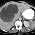
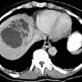
RADIOLOGY: HEPATOBILIARY: Case# 32863: HEPATIC ABSCESS. A 76-year-old black female with right upper quadrant pain and new onset of leukocytosis (31,000) without fever. There is a 14.0 x 9.0 x 10.0 cm low attenuation mass with minimal peripheral enhancement and thick septations associated with its superior aspect. There is minimal associated biliary dilatation. Pyogenic liver abscesses occur predominantly in individuals with other underlying disorders, most commonly biliary tract disease. Abscesses may also occur following hepatic transplantation. Clinical findings are nonspecific with fever and jaundice being the most common. Diagnosis is best made by contrast-enhanced CT or ultrasound. Pyogenic abscesses are usually solitary and often multiloculated with thickened enhancing walls. Gas is present in about 20% of cases. Amoebic abscesses are also solitary lesions. Pyogenic abscesses may be distinguished from amoebic abscesses on the basis of clinical findings. Whereas patients with pyogenic abscesses are usually septic, patients with amoebic abscesses are not, and they usually have a history of travel to an endemic area. Pyogenic abscesses require drainage while amoebic abscesses do not and may be treated with antibiotics.
- Author
- Peter Anderson
- Posted on
- Thursday 1 August 2013
- Tags
- hepatobiliary, radiology
- Albums
- Visits
- 728
0 comments