)
)

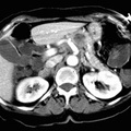
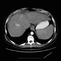
RADIOLOGY: ABDOMEN: Case# 32813: ?HCC. This 41 year old male had recent angiography demonstrating a spoke wheel 1.5 cm lesion in the medial aspect of the left lower pole kidney as well as atypical vasculature in the liver. 1. Multiple enhancing hepatic masses suspicious for hepatocellular carcinoma. Correlation with AFP would be helpful. 2. Enhancing ill-defined left renal mass. The differential would include renal cell carcinoma, oncocytoma, as well as lymphoma. Metastatic disease from hepatocellular carcinoma would be much less likely. The lesion appears well contained within the kidney and there is no pathological lymphadenopathy. These findings suggest that the hepatic lesions are not metastases from the renal lesion. 3. Wedged shaped opacity in the left lung base. This may represent pneumonia. Alternatively, does the patient have any clinical evidence of pulmonary embolism?.
- Author
- Peter Anderson
- Posted on
- Thursday 1 August 2013
- Albums
- Visits
- 1174
0 comments