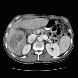
RADIOLOGY: HEPATOBILIARY: Case# 32808: F/U HCC WITH BLEED. The patient is a 54 year old male. 10/11/95:1) The previously described low attenuation lesion in the posterior segment of the right hepatic lobe has greatly increased in size since 8/30/93 and now involves all segments of the liver. The appearance is consistent with diffuse hepatocellular carcinoma. 2) Large right sacral metastases (as described above). A second metastatic lesion is suspected in the body of the L5 vertebra (see above). 3) A 1.5 cm low attenuation lesion is seen in the upper pole of the left kidney. Differential diagnosis would include primary tumor versus metastasis or complicated cyst. 4) Multiple intra-abdominal varices, with a splenorenal shunt. 5) Cholelithiasis. ABDOMINAL CT 11/3/95: 1) Large amount of free intraperitoneal fluid with increased attenuation consistent with acute hemorrhage. Probable active bleeding is demonstrated into the fluid surrounding the liver. The hemoperitoneum is seen around the liver and spleen with extension into the pelvis. 2) Diffuse hepatocellular carcinoma with involvement of all segments of the liver. The previously described low attenuation lesion in the right hepatic lobe is unchanged in size from the previous examination. However, this lesion demonstrates a marked decrease in attenuation since the previous examination which may be consistent with infarction. The predominant lesion in the left hepatic lobe has increased in size from 3 x 4 cm to 5 x 4 cm. 3) Multiple 1 cm nodules are seen in the right and left lung bases which most likely represent metastatic disease. 4) Interval increase in the size of the right sacral soft tissue mass causing bone destruction, as described above. There is also bone destruction of the ischial bones bilaterally. 5) The previously described 1.5 cm low attenuation lesion in the upper pole of the left kidney is unchanged since the prior examination. 6) The spleen has decreased in size from the prior examination. The spleen previously measured 14.5 cm and now measures 10.5 cm craniocaudal. 7) Multiple intraabdominal varices with a surgical splenorenal shunt.