)
)

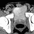
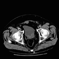
RADIOLOGY: ABDOMEN: Case# 32799: INGUINAL HERNIA. Patient is a 36 year old male referred for evaluation of abnormal renal ultrasound with possible left renal mass. The left kidney is long and has duplication of the collecting system down at least to the pelvic rim where the two ureters appear to join. There is no evidence of mass or focal lesion. The right kidney and both adrenal glands are within normal limits in appearance. There is large right sided inguinal hernia with a loop of small bowel and some fluid within it. There is no evidence of obstruction or strangulation at this time. While most hernias may be diagnosed clinically, CT may be useful in differentiating between a hernia and a mass within the abdominal cavity or wall. CT may also identify incisional hernias in patients undergoing postoperative evaluation. The inguinal hernia, which is caused by herniation of peritoneal contents through the deep inguinal ring, is the most common type of hernia. Large inguinal hernias may extend into the labium majus or scrotum. CT may demonstrate bowel or omentum within a hernia sac and narrowing of gas-filled small intestine and mesenteric fat and vessels as they pass through the peritoneal defect.
- Author
- Peter Anderson
- Posted on
- Thursday 1 August 2013
- Albums
- Visits
- 1121
0 comments