)
)

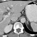
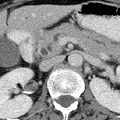
RADIOLOGY: PANCREAS: Case# 32787: PANCREATIC MASS-ADENOCARCINOMA. This is a 47 year old white female without significant prior medical history, who presents with three weeks of nausea with new onset of jaundice. There is intrahepatic biliary and pancreatic duct dilatation. An enhancing 3 x 3 x 4 cm pancreatic head mass is identified. The mass abuts the right lateral surface of the superior mesenteric vein, but there is apparent preservation of a separating fat plane. The superior mesenteric artery, portal vein, and IVC are patent and show no evidence of encasement. A single 12 mm lymph node is identified within the porta. No other retroperitoneal adenopathy is seen. In the lateral segment of the left hepatic lobe is a 7mm hypodense lesion which is suspicious for metastatic disease. An ill defined low density lesion may also be present in the posterior segment of the right hepatic lobe. The gastrohepatic ligament is free of adenopathy. This almost uniformly fatal cancer is the 4th most common malignant tumor accounting for 5% of cancer deaths in the United States. Symptoms are usually nonspecific and insidious such that the cancer is advanced by the time of diagnosis. Most cancers occur in the head of the pancreas and are usually adenocarcinomas arising from ductal cells. The sensitivity of CT scan in diagnosing pancreatic carcinoma is approximately 95% whereas that of ultrasound is less than 80% for cancer of the head and 40% for the tail. The primary finding on CT is a focal mass;however, if no mass is present, other findings may suggest neoplasm. First, the pancreas may become more heterogeneous in density with age. Thus, a focal region of homogeneous soft tissue density might raise suspicion of a carcinoma. The presence of both a dilated common bile duct and a dilated main pancreatic duct in the absence of calculus suggests ampullary or pancreatic head neoplasm, but this may also be seen in benign disease. The finding of a dilated main pancreatic duct in the body or tail but not in the head or neck suggests neoplasm, and finally, the finding of rounded convex borders of both anterior and posterior surfaces of the uncinate process raises suspicion for carcinoma. Ten to fifteen percent of patients will have potentially resectable tumors, and they may be distinguished by CT findings. Signs of potential resectability include an isolated pancreatic mass with or without dilatation of the bile and pancreatic ducts and combined bile-pancreatic duct dilatation without an identifiable pancreatic mass. Extension of the tumor beyond the margins of the pancreas, involvement of adjacent organs, liver metastases, ascites, and regional adenopathy are signs of unresectability. Unfortunately, most of those who undergo resection eventually die of pancreatic cancer.
- Author
- Peter Anderson
- Posted on
- Thursday 1 August 2013
- Albums
- Visits
- 1052
0 comments