)
)
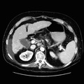
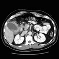
 Gallbladder wall thickening, gallbladder sludge and possibly cholelithiasis, mucosal irregularity, and severe strandy change surrounding the gallbladder which all indicate cholecystitis and probably gallbladder necrosis in this patient with right upper quadrant pain and fever. 2) Inflammatory change in adjacent hepatic flexure with no evidence of bowel obstruction. 3) Bibasilar atelectasis/infiltrate with bilateral small pleural effusions. Infection in these areas cannot be excluded and recommend correlation clinically and with recent plain films of the chest. Acute cholecystitis, or inflammation of the gallbladder, can be classified as calculous (associated with gallstones) or acalculous. Gangrenous cholecystitis is a rara form of acute cholecystitis which is often indistinguishable from acute cholecystitis. The mortality rate of gangrenous cholecystitis is 22% while the mortality rate for acute cholecystitis is only around 1 to 6%. Symptoms associated with the onset of acute cholecystitis include progressive right upper quadrant or epigastric pain, mild fever, anorexia, tachycardia, diaphoresis, and nausea and vomiting. Lab abnormalities associated with this condition include mild to moderate leukocytosis often accompanied by mild elevations in serum alkaline phosphatase values. Most cases of acute cholecystitis are diagnosed clinically, via ultrasound, or via radionuclide hepatobiliary scan. CT is used to evaluate complicated or atypical cases. CT findings include gallstones (in 95% of patients), distended gallbladder lumen (greater than 5cm), thickening of the gallbladder wall (greater than 3mm), halo of subserosal edema in the gallbladder wall, pericholecystic fluid collection associated with perforation, increase in bile density (20 H) (due to biliary stasis, intraluminal pus, hemorrhage. or cellular debris), and air in the gallbladder wall (emphysematous cholcystitis).")
RADIOLOGY: HEPATOBILIARY: Case# 32769: CHOLECYSTITIS. 58 year old male with right upper quadrant pain, fever, and elevated bilirubin. 1) Gallbladder wall thickening, gallbladder sludge and possibly cholelithiasis, mucosal irregularity, and severe strandy change surrounding the gallbladder which all indicate cholecystitis and probably gallbladder necrosis in this patient with right upper quadrant pain and fever. 2) Inflammatory change in adjacent hepatic flexure with no evidence of bowel obstruction. 3) Bibasilar atelectasis/infiltrate with bilateral small pleural effusions. Infection in these areas cannot be excluded and recommend correlation clinically and with recent plain films of the chest. Acute cholecystitis, or inflammation of the gallbladder, can be classified as calculous (associated with gallstones) or acalculous. Gangrenous cholecystitis is a rara form of acute cholecystitis which is often indistinguishable from acute cholecystitis. The mortality rate of gangrenous cholecystitis is 22% while the mortality rate for acute cholecystitis is only around 1 to 6%. Symptoms associated with the onset of acute cholecystitis include progressive right upper quadrant or epigastric pain, mild fever, anorexia, tachycardia, diaphoresis, and nausea and vomiting. Lab abnormalities associated with this condition include mild to moderate leukocytosis often accompanied by mild elevations in serum alkaline phosphatase values. Most cases of acute cholecystitis are diagnosed clinically, via ultrasound, or via radionuclide hepatobiliary scan. CT is used to evaluate complicated or atypical cases. CT findings include gallstones (in 95% of patients), distended gallbladder lumen (greater than 5cm), thickening of the gallbladder wall (greater than 3mm), halo of subserosal edema in the gallbladder wall, pericholecystic fluid collection associated with perforation, increase in bile density (20 H) (due to biliary stasis, intraluminal pus, hemorrhage. or cellular debris), and air in the gallbladder wall (emphysematous cholcystitis).
- Author
- Peter Anderson
- Posted on
- Thursday 1 August 2013
- Tags
- hepatobiliary, radiology
- Albums
- Visits
- 845
0 comments