)
)

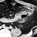
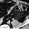
RADIOLOGY: GASTROINTESTINAL: GI: Case# 114: DIVERTICULITIS WITH PERFORATION. This is a 62 year old man with a large amount of free intraperitoneal air identified on plain films. There is a large volume of free intraperitoneal air. An air fluid layer is noted anteriorly. However, there are also several small bubbles of gas located throughout the mesentery. A large inflammatory area is seen in the left lower quadrant in the region of the proximal sigmoid. However, no loculated or drainable fluid collection is seen in this location. These findings are consistent with a proximal sigmoid diverticulitis with perforation. There may be some involvement of nearby small bowel in the inflammatory process. A region of localized infection involving diverticula may lead to microabscess formation resulting in diverticulitis. Diverticulitis may be complicated by perforation, fistulas, and large abscesses. A frank abscess may be seen on CT in about one-third of patients with diverticulitis. This will appear as an extraluminal mass of soft-tissue density and a low-density center that may contain gas or, if in communication with the lumen, contrast. Thickening of the bowel wall is also evident. Abdominal films may show collections of air and fluid in the left lower quadrant. Large abscesses, fistulas, and perforations require surgical treatment.
- Author
- Peter Anderson
- Posted on
- Thursday 1 August 2013
- Tags
- gastrointestinal, radiology
- Albums
- Visits
- 995
0 comments