)
)

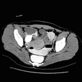
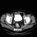
RADIOLOGY: GASTROINTESTINAL: GI: Case# 111: RECTAL CA. Patient is a 70 year old male. 1. 7 to 8cm length of irregular nearly circumferential wall thickening at the rectosigmoid junction is most consistent with carcinoma. There are no definite nodes or metastases but there is some slight strandiness of the perirectal fat which is worrisome for invasion. 2. Unusual liver contour with atrophy of the lateral segment of the left lobe. Multi-loculated fluid density lesion along that margin of the liver probably represents atrophied left lobe with small dilated ducts. The etiology of this is uncertain but the remainder of the liver is normal in appearance. 3. Multiple renal cysts, some quite large as described above. All are simple without evidence of tumor. 4. Aorto-iliac atherosclerosis.
- Author
- Peter Anderson
- Posted on
- Thursday 1 August 2013
- Tags
- gastrointestinal, radiology
- Albums
- Visits
- 988
0 comments