)
)
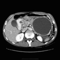
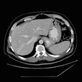
. A pseudocyst may have a persistent connection to the pancreatic duct which may be demonstrated by ERCP. Typically, an uncomplicated pseudocyst will have a homogeneous water density. Increased density or inhomogeneity indicate some complicating factor such as hemorrhage or infection. Psuedocysts may also form in the walls of bowel (stomach, duodenum, colon) due to extension of pancreatic enzymes into the surrounding tissues.")
RADIOLOGY: PANCREAS: Case# 101: LARGE PANCREATIC PSEUDOCYST. The patient is a 48 year old male with history of pancreatitis with a large LUQ cystic structure seen on ultrasound. A 12 x 12 x 13 cm fluid collection is seen in the region of the pancreatic tail immediately inferior to the greater curvature of the stomach displacing it superiorly. This fluid collection demonstrates peripheral calcifications. A second fluid collection is seen measuring 2 x 1.5 cm with septations, and is present in the region of the pancreatic head. The pancreatic duct is enlarged measuring 1 cm, with calcifications within the duct. The pancreas itself is irregular and demonstrates multiple calcifications especially at the head and is consistent with chronic pancreatitis. Pancreatic pseudocysts are usually the sequelae of pancreatitis, most often associated with the subacute or chronic varieties. Peripancreatic fluid collections in the setting of acute pancreatitis are not pseduocysts. A pseudocyst is a cystic structure with a thick, fibrous wall and is not lined with epithelium (hence, it is not a true cyst). A pseudocyst may have a persistent connection to the pancreatic duct which may be demonstrated by ERCP. Typically, an uncomplicated pseudocyst will have a homogeneous water density. Increased density or inhomogeneity indicate some complicating factor such as hemorrhage or infection. Psuedocysts may also form in the walls of bowel (stomach, duodenum, colon) due to extension of pancreatic enzymes into the surrounding tissues.
- Author
- Peter Anderson
- Posted on
- Thursday 1 August 2013
- Albums
- Visits
- 1117
0 comments