)
)

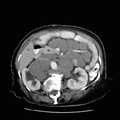
RADIOLOGY: ABDOMEN: Case# 89: LYMPHOMA. 68-year-old woman with abdominal mass on physical examination. There is extensive adenopathy present throughout the retroperitoneum, tracking into the mesentery and porta hepatis. The right kidney is displaced laterally. There is moderate right hydronephrosis caused by the adenopathy. The adenopathy tracts into the pelvis along the iliac vessels. The adenopathy displaces bowel, the pancreas and the great vessels of the abdomen. Computed tomogaphy has, to a large extent, replaced lymphangiography in the evaluation of lymphoma. CT can demonstrate retroperitoneal and pelvic nodal enlargement with virtually the same sensitivty as lymphangiography; however, CT depicts adenopathy in locations that lymphangiography cannot: around the celiac axis, retrocrural space, renal/splenic/hepatic hila, and the mesentery. CT also demonstrates lymphomatous involvement of other organs, such as the kidneys, spleen, liver and GI tract.
- Author
- Peter Anderson
- Posted on
- Thursday 1 August 2013
- Albums
- Visits
- 1546
0 comments