Difference between revisions of "File:IPLab2Atrophy5.jpg"
(This is a higher-power photomicrograph indicating loss of testicular parenchymal tissue. There are very few recognizable spermatic cells in this tissue. The cluster of cells in the upper right is a focus of interstitial or Leydig cells (arrow). These c...) |
(Peter Anderson uploaded a new version of "File:IPLab2Atrophy5.jpg": A seminiferous tubule is shown on the left containing remnants of spermatocytes (1). There are no mature sperm present. On the right-hand portion of the slide are remnants o) |
(No difference)
| |
Latest revision as of 16:04, 19 August 2013
This is a higher-power photomicrograph indicating loss of testicular parenchymal tissue. There are very few recognizable spermatic cells in this tissue. The cluster of cells in the upper right is a focus of interstitial or Leydig cells (arrow). These cells are not affected by the hormone-induced atrophy process.
File history
Click on a date/time to view the file as it appeared at that time.
| Date/Time | Thumbnail | Dimensions | User | Comment | |
|---|---|---|---|---|---|
| current | 16:04, 19 August 2013 | 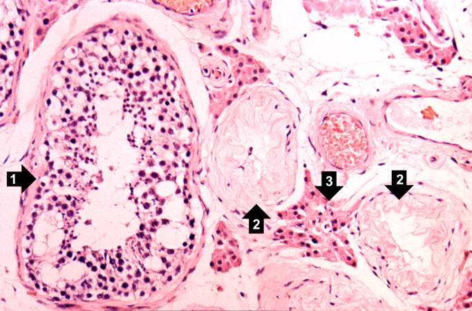 | 683 × 450 (60 KB) | Peter Anderson (talk | contribs) | A seminiferous tubule is shown on the left containing remnants of spermatocytes (1). There are no mature sperm present. On the right-hand portion of the slide are remnants of other spermatic tubules which have completely atrophied and lost all of their... |
- You cannot overwrite this file.
File usage
The following page links to this file: